Homepage |  Send us an Email | Legal Notice |  German Homepage   English Homepage


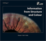
Information from Structure and Colour

Iridology 1

The superb quality of this unique and beautiful text- and reference book is not only enhanced by the more than 30 years of practical experiences by each of the three authors but also by 180 color illustrations (including approx. 120 excellent iris photos in color).

On 264 pages, Willy Hauser, Josef Karl, and Rudolf Stolz explain the topography, constitutions - disposition - diathesis, structure markings, and pigments of the eye in an easily understandable way, based on the new regulations of 1996-1997.


Specifications:
264 pages, 10“ x 9“, high-quality art paper, special sturdy dirt-repelling binding,
hard cover, all pages sealed with protective varnish. Printed in Germany.


€ 98,- plus shipping costs per book:
€ 21 within postal Europe / € 34 outside postal Europe).

Information_from_Structure_and_Colour.pdf

Discription of the book.

Typ: pdf - Größe: 73.7 KB - Aktualisierung: 05.10.2010 - Downloads: 7768

Content

  • 1. History of Eye Diagnosis

  • 2. Constitution-Disposition-Diathesis:
    • What do we mean by constitution?
    • What do we mean by disposition (type - structural)?
    • What do we mean by diathesis (regulatory)?

    • Constitutions:
      • Lymphatic constitution.
      • Hematogenic constitution.
      • Mixed constitution.

    • Dispositions:
      • Neurogenic type.
      • Weak mesenchyme type.
      • Vegetative-spastic type.
      • Weak glandular type.
      • Tuberculine type.

    • Diatheses:
      • Exsudative diathesis (hydrogenoid).
      • Uric acid diathesis.
      • Lipemic diathesis.
      • Allergic diatheses

  • 3. Structural signs:
    • Lacunae:
      • Closed lacuna (simple, genetic inhirited).
      • Structured leaf-rip lacuna.
      • Closed lacuna with complicatin signs.
      • Closed lacuna with surrounding bright radials.
      • Open lacuna.
      • Single sided lacuna.
      • Asparagus formed lacuna.
      • Torpedo- and lancet lacuna.
      • Giant lacuna.
      • Ladder- and rung lacuna.
    • Honeycombs.
    • Crypts.
    • Substance defects.
    • Transversales as genotypes.
    • Neuronic networks.
    • Corkscrew radials.
    • Weak mesenchyme disposition (connective tissue weakness-iris).
    • Weak glandular disposition (daisy or gland iris).
    • Spasm rings, spastic furrows and radii solaris.
    • Perifocal markings.
    • Stages of stroma.

  • 4. Pigments:
    • Introduction.
    • Criteria of evaluation.
    • Analysis of single pigments.
    • Urorosein pigment.
    • Gastrin pigment.
    • Orange-colored pigments.
    • Hepatotrophic pigments.
    • Basics about pigments.
    • Missing alien pigments.
    • Multitude of hepatotrophic pigments.
    • Single pigments (solitary pigments).
    • The sectorial heterochromia.
    • Color changes in the wreath zone.
    • The phenomenon of the accompanying shadow.
    • Bright wreath zone.
    • Darkened wreath zone.
    • Complete heterochromia.
    • Different pigmentations.
    • Pigments in the ruffle zone.

  • 5. Topography:
    • Preliminary remarks.
    • Why the term topography?
    • Gastric-intestinal zone.
    • The intestinal tract.
    • The pancreas.
    • The liver and the biliary system.
    • The urogenital system.
    • The pulmo-bronchus, pleura, cor and mamma.
    • The nasal sectors.
    • The topography of the thyroid.
    • The topography of the ear and the occiput.
    • The topography of the brain.
    • Possibilities and limits.
    • Final remarks.
    • Index.
    • Bibliography
top of page | back to previous page | Show 'printer friendly' page